

Alopecia Areata Treatment Case Study – 6-Month Hair Regrowth Journey with Kenacort Injection

Alopecia Areata Treatment Case Study – 6-Month Hair Regrowth Journey with Kenacort Injection

Written by: Dr. Kiran Chotaliya Editorial Team  ·  Reviewed by: Dr. Kiran Chotaliya, M.D. Dermatologist & Chief Hair transplant Surgeon  ·  Hairfree & Hairgrow Clinics, India  ·  Updated Oct 2026

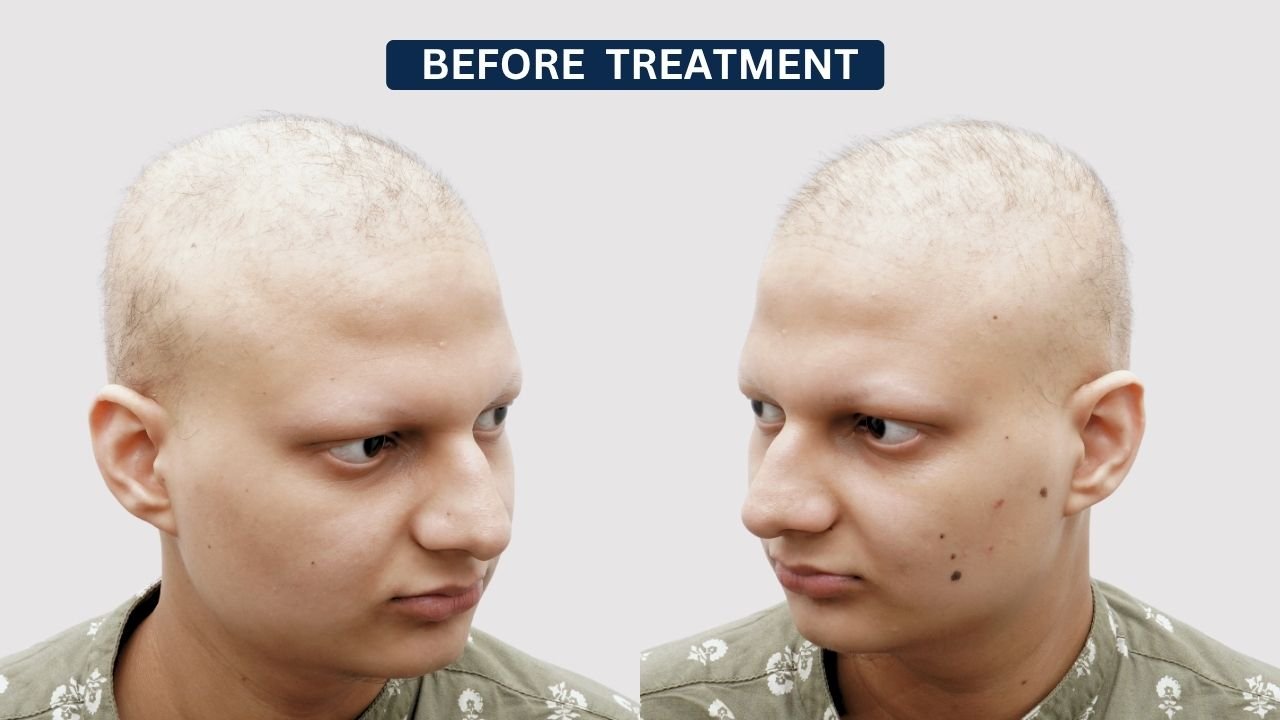
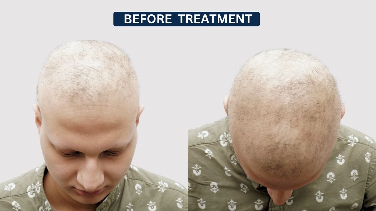
This case study documents the 6-month treatment journey of a 19-year-old male patient diagnosed with alopecia areata. Before treatment, the patient presented with extensive scalp hair loss and very limited visible hair coverage, with significant scalp exposure.

According to the provided patient profile, there was no significant medical history, and the treatment plan included Kenacort Injection for alopecia areata. Serial photographs document visible changes at approximately 1, 3, 5, and 6 months.

The case study below follows the same detailed clinical format as the reference document, including patient profile, clinical assessment, treatment plan, month-wise progress, outcome, aftercare, disclaimer, and FAQs. Pasted markdown

Patient Consent Notice

This case study has been published with the patient’s informed consent for educational and informational purposes. Appropriate measures have been taken to maintain patient privacy while presenting the scalp condition, treatment approach, and documented clinical progress.

Individual outcomes with alopecia areata treatment can vary. Hair regrowth may depend on disease activity, extent of hair loss, duration of the condition, individual response to treatment, recurrence, and other biological factors.

The result shown in this case should not be considered a guarantee of similar outcomes for every patient.

Patient Case Overview

Patient Details

Information

Age

19 years

Gender

Male

Diagnosis

Alopecia Areata

Medical History

No sgniificant medical history reported

Initial Condition

Extensive scalp hair loss with marked scalp exposure

Affected Area

Scalp

Treatment Plan

Kenacort Injection

Treatment Type

Intralesional corticosteroid treatment

Documented Follow-Up

1, 3, 5 and 6 months

Latest Documented Progress

6 months


NAMISH ARORA ALOPECIA

Case Overview

Age, City

47 Years,
Pune, India

Hair Loss Duration

12–15 Years

Baldness Grade

Norwood
Grade 4–5

Areas Affected

Hairline,
Mid Scalp,
Crown Thinning

Techniques

Big FUE

Grafts

3220+

Patient’s Main Concern

The patient presented with extensive scalp hair loss, resulting in marked scalp visibility and very limited overall hair coverage.

The pretreatment photographs show that much of the scalp was exposed, with only short and sparse residual hair visible in some areas.

His main concerns included:

  • Extensive scalp hair loss
  • Significant scalp visibility
  • Very low overall hair density
  • Uneven hair distribution
  • Difficulty concealing the affected scalp
  • Cosmetic concern related to visible hair loss
  • Desire to regain scalp hair coverage
  • Concern about continued or recurrent hair loss

Clinical Assessment

The patient’s alopecia areata was assessed based on the location, appearance, and extent of the localized patches.

Clinical evaluation focused on:

  • Number of alopecia patches
  • Location of affected areas
  • Degree of visible scalp exposure
  • Existing hair around the patches
  • Evidence of developing regrowth
  • Response to treatment at follow-up
  • Presence of new or recurrent patches

Serial photographs were used to document the visible treatment response over a five-month period. Pasted markdown

Initial Alopecia Areata Condition

Before starting treatment, the patient’s clinical photographs showed marked scalp hair loss with only minimal visible hair remaining.

The baseline condition included:

  • Significant scalp exposure
  • Very sparse visible hair
  • Reduced hair density across a large portion of the scalp
  • Limited frontal and upper scalp coverage
  • Uneven distribution of remaining hair
  • Extensive visible involvement rather than a small isolated patch

This baseline served as the reference point for evaluating subsequent treatment progress.

Detailed Graft Breakdown for NW3 Restoration

Single Hair Grafts
530
For Natural Hairline
Multi Hair Grafts
2480
Mid Scalp Coverage
Total Grafts Done
3010
Total Grafts Transplanted: (add case specific number)
Parameter
No. of Grafts
Details
Single hair grafts
530
For natural hairline
Multi hair grafts
2480
Mid scalp coverage
Total grafts done
3010

Clinical Assessment

The patient was assessed clinically for alopecia areata according to the extent and distribution of scalp hair loss.

The clinical evaluation focused on:

  • Extent of scalp involvement
  • Amount of remaining hair
  • Degree of visible scalp exposure
  • Distribution of hair loss
  • Appearance of new hair during follow-up
  • Changes in hair density
  • Improvement in scalp coverage
  • Response to treatment
  • Appearance of any new areas of hair loss

Serial photographs were used during follow-up to document visible changes, consistent with the photographic monitoring approach used in the supplied reference case-study format. Pasted markdown

Personalized Alopecia Areata Treatment Plan

Following clinical evaluation, the patient was advised Kenacort Injection treatment for alopecia areata.

Kenacort contains triamcinolone, a corticosteroid that may be administered intralesionally by dermatologists in selected cases of alopecia areata. The amount, concentration, injection sites, treatment interval, and total number of sessions should be determined according to the treating dermatologist’s assessment and individual patient response. Pasted markdown

Treatment Goals

The treatment plan focused on:

  • Managing alopecia areata
  • Encouraging visible hair regrowth
  • Improving overall scalp coverage
  • Reducing scalp visibility
  • Supporting progressive maturation of new hair
  • Monitoring treatment response
  • Checking for recurrence or new hair-loss areas
  • Maintaining regular clinical follow-up

Kenacort Injection Treatment Overview

Kenacort Injection was used according to the dermatologist’s treatment plan.

Treatment was directed toward affected scalp areas based on the clinical presentation.

Follow-up assessments focused on:

  • Development of new hair
  • Improvement in hair density
  • Reduction in scalp exposure
  • Distribution of regrowing hair
  • Hair thickness and maturation
  • Appearance of new alopecia areas
  • Stability of previously improved areas
  • Need for further treatment

The photographic sequence indicates that regrowth developed gradually over several months rather than immediately.

Month-by-Month Alopecia Areata Treatment Progress

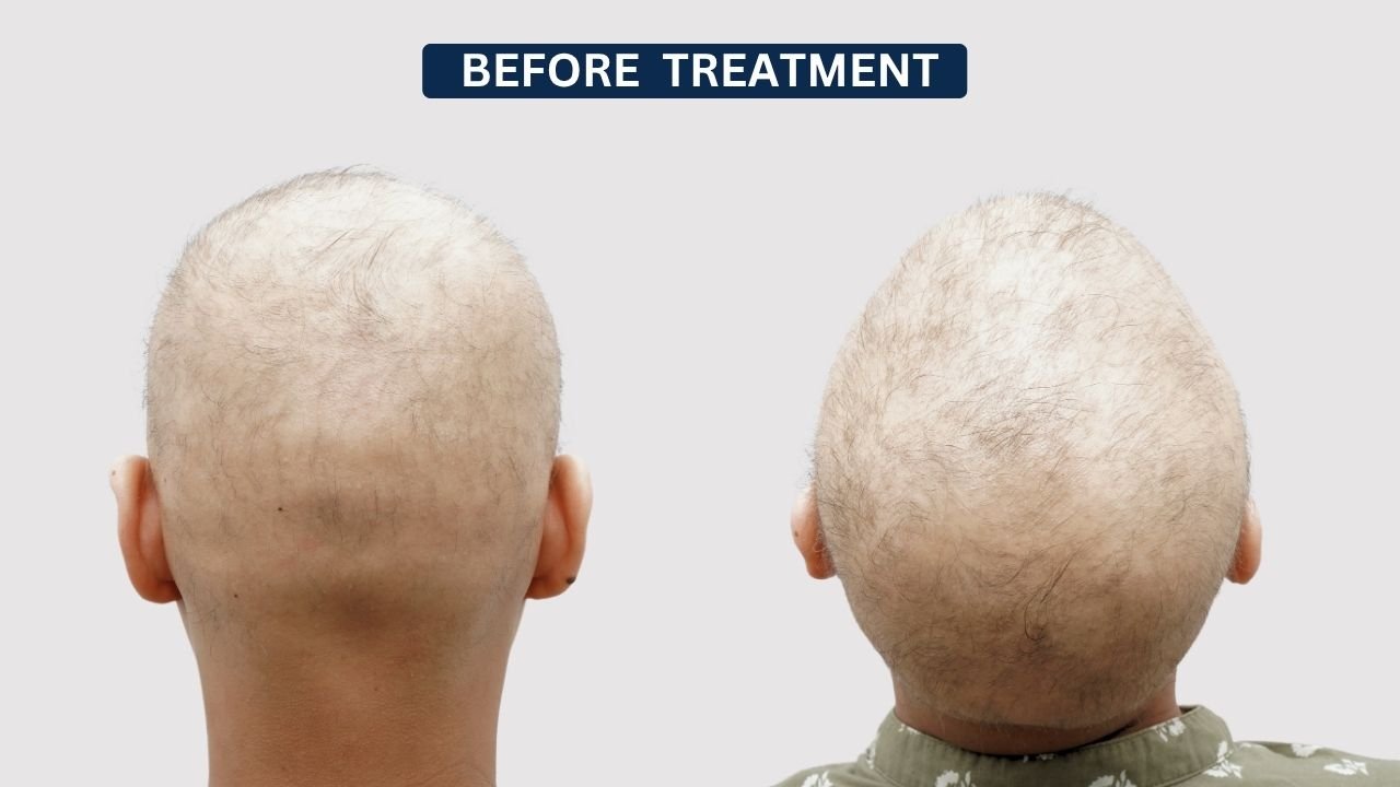
Before Treatment – Baseline Condition

At baseline, the patient showed extensive scalp hair loss with substantial scalp exposure.

The photographs demonstrated:

  • Very sparse scalp hair
  • Marked reduction in overall density
  • Extensive visible scalp
  • Minimal coverage across the frontal and upper scalp
  • Short, fine residual hair in some areas

This stage served as the baseline for later comparison.

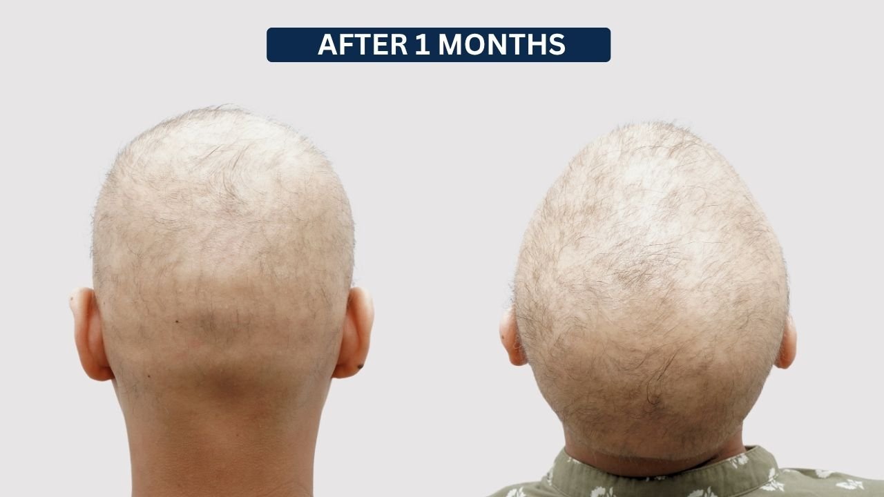
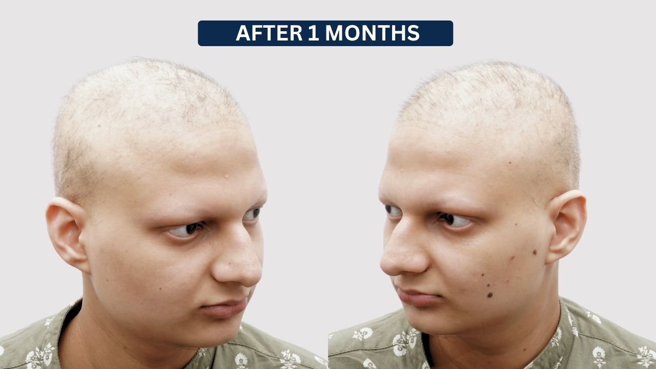
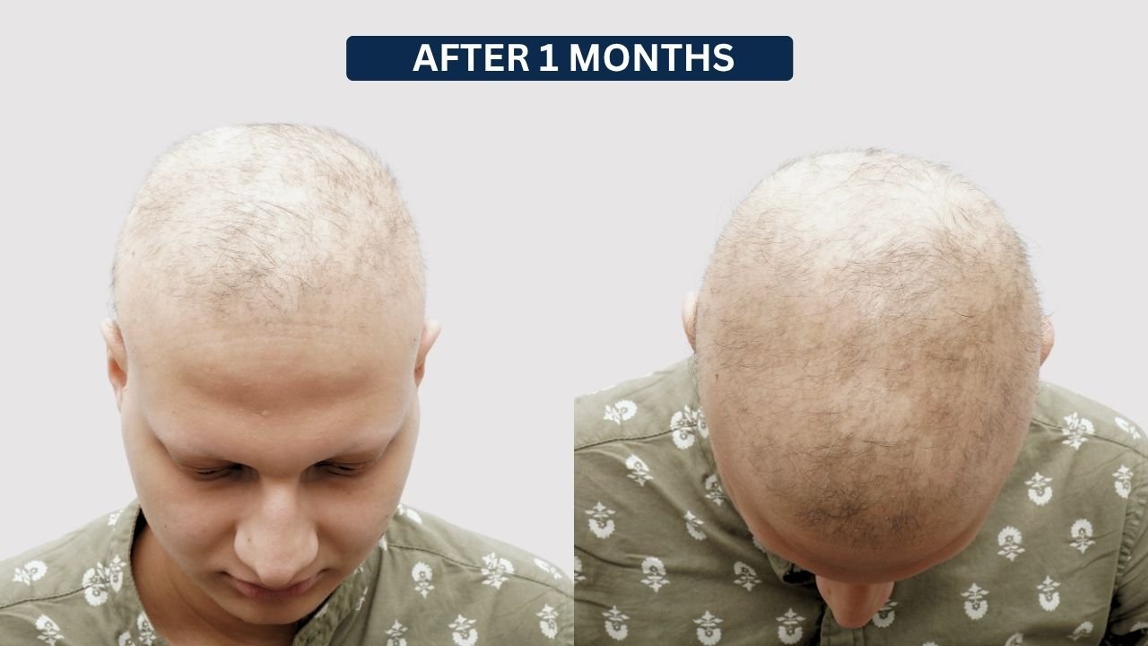
After 1 Month – Early Treatment Response

At approximately 1 month, the scalp remained highly visible and overall density was still very limited.

However, the photographs showed some short and fine hair across parts of the scalp.

Observed features included:

  • Persistent extensive scalp exposure
  • Fine short hairs visible over portions of the scalp
  • Limited overall density
  • Early developing regrowth
  • Minimal change in cosmetic coverage at this stage

This represented a very early treatment stage and not the final outcome.

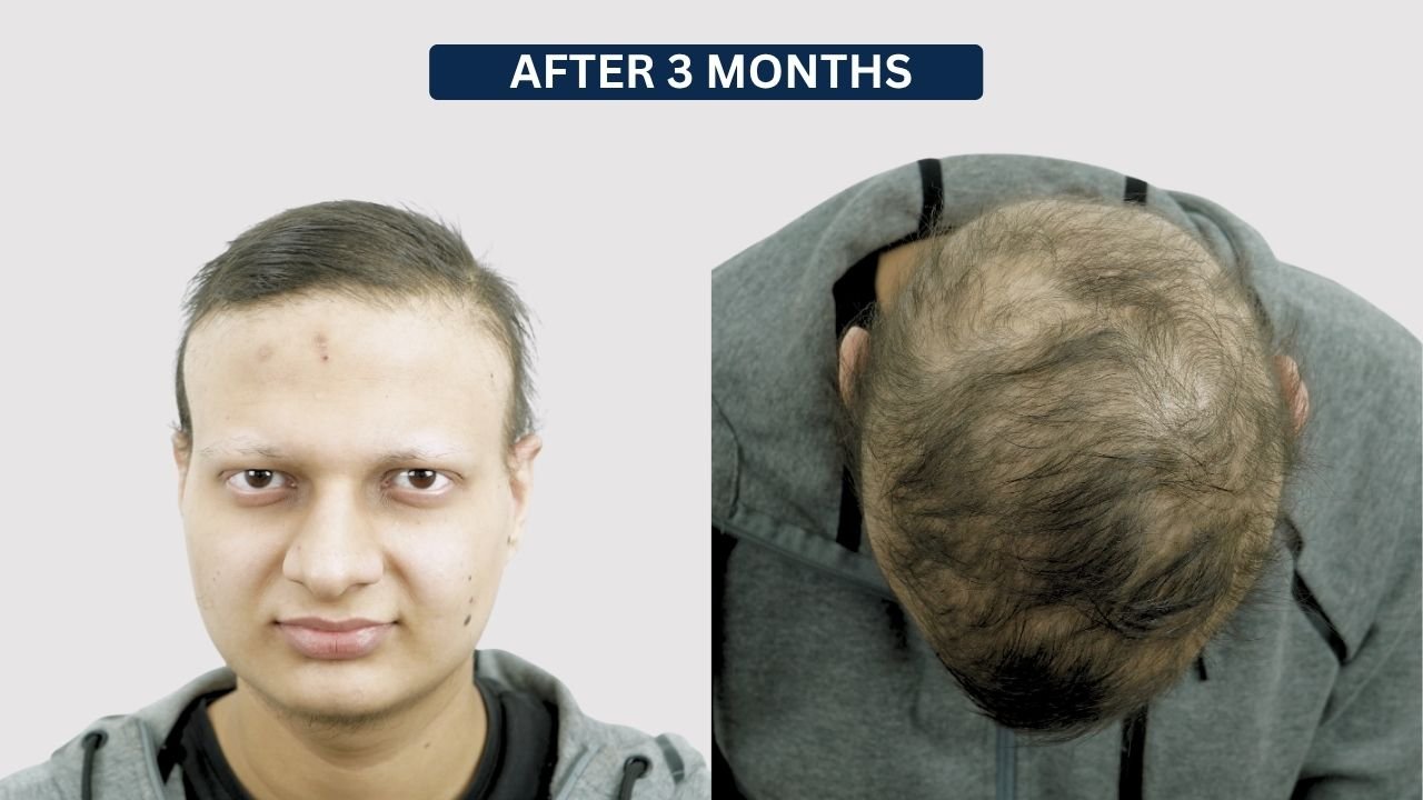
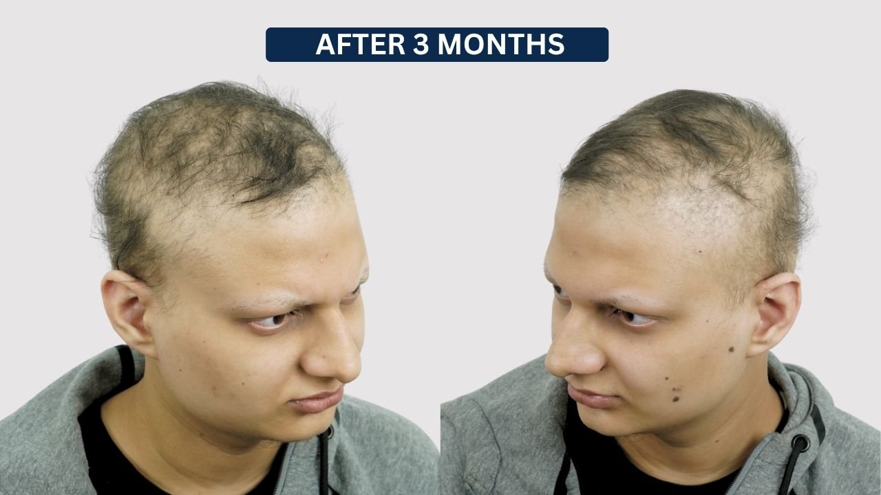
After 3 Months – Developing Regrowth

At the 3-month follow-up, considerably more hair was visible across the scalp compared with baseline.

The photographs showed:

  • Increasing short hair growth
  • Improved distribution of hair across the scalp
  • Reduced completely bare appearance
  • Better frontal and upper scalp coverage
  • Continued visible thinning in several regions
  • Persistent scalp exposure through the developing hair

At this stage, the hair was still relatively fine and coverage remained incomplete.

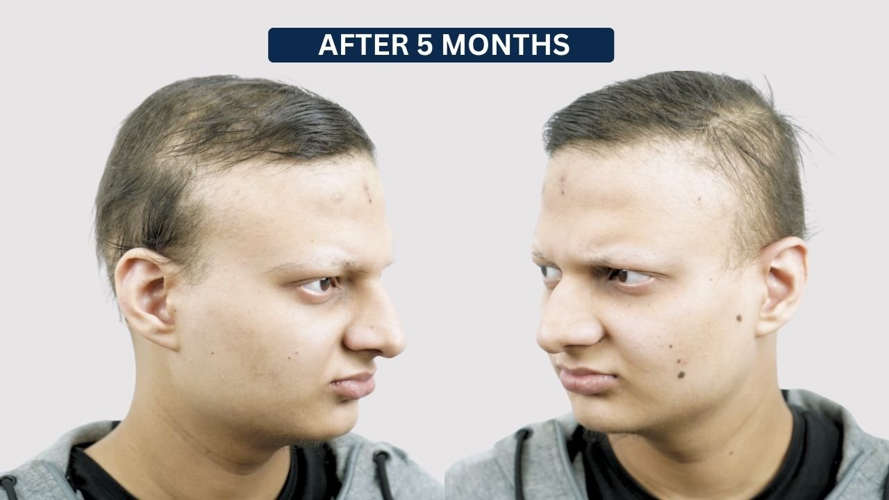
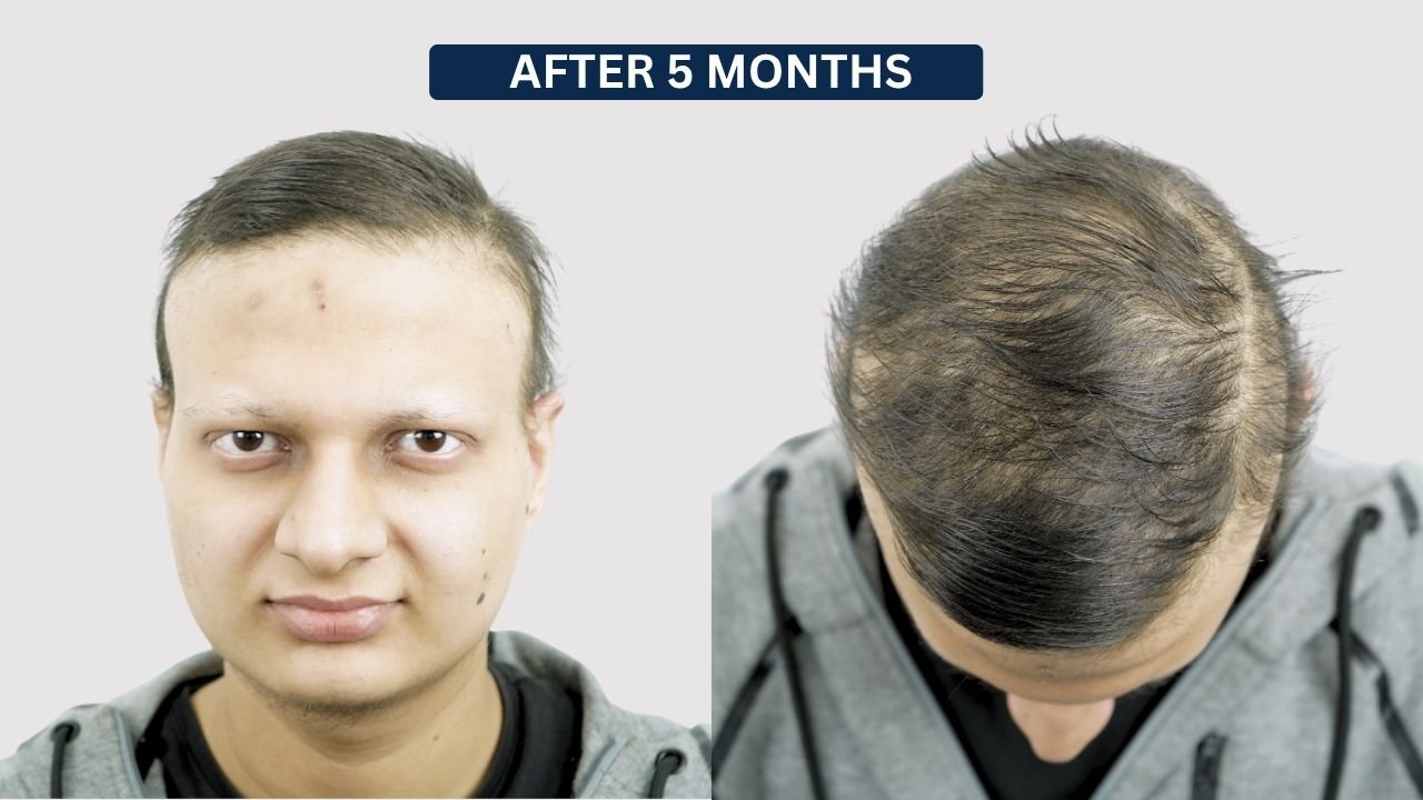
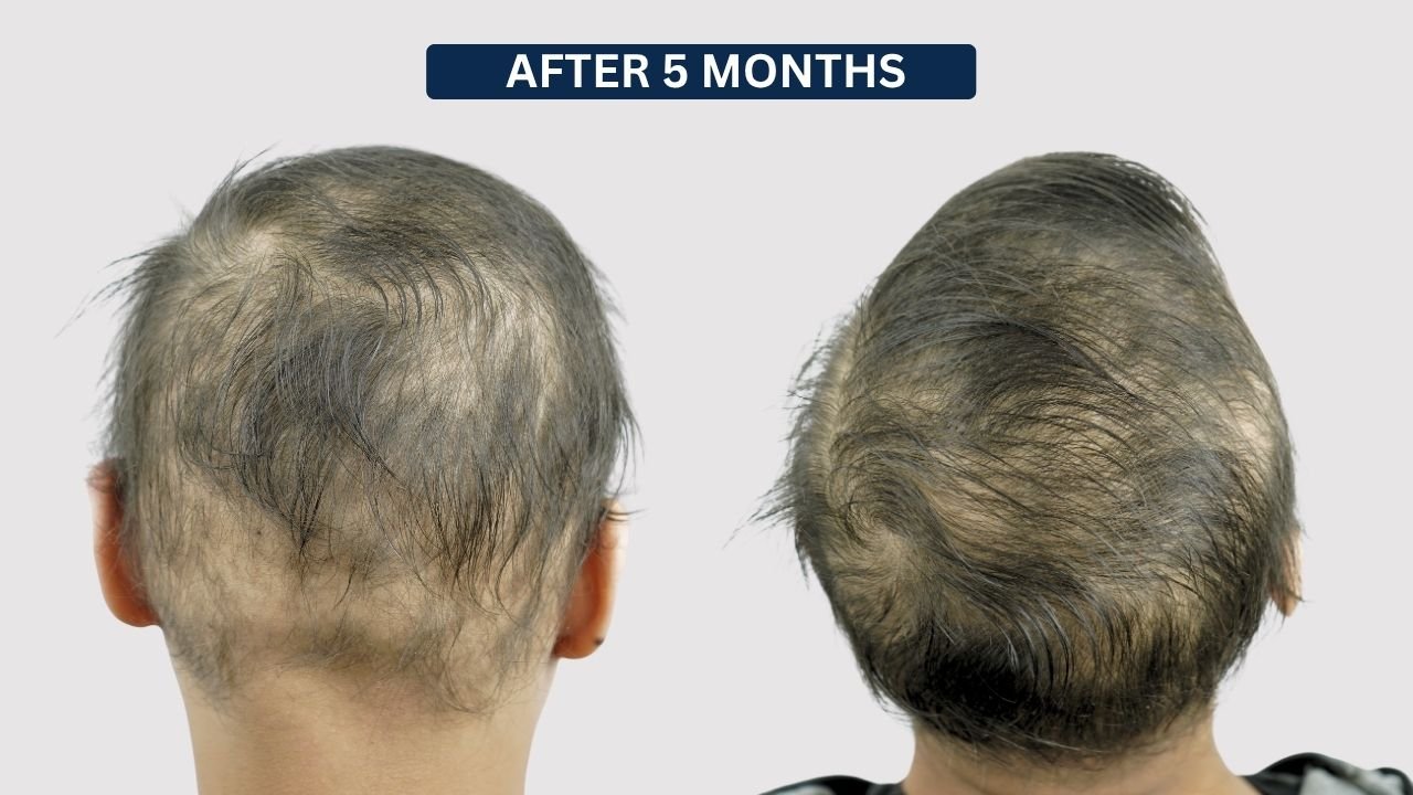
After 5 Months – Progressive Hair Coverage

By approximately 5 months, the patient showed further visible improvement.

Compared with the 3-month stage:

  • Hair had become longer
  • Overall scalp coverage had improved
  • Frontal hair appeared more established
  • More hair was visible through the upper scalp
  • Areas of complete scalp exposure were reduced
  • Density remained lower across some central and crown regions

The 5-month photographs indicate ongoing regrowth and maturation, although visible thinning was still present.

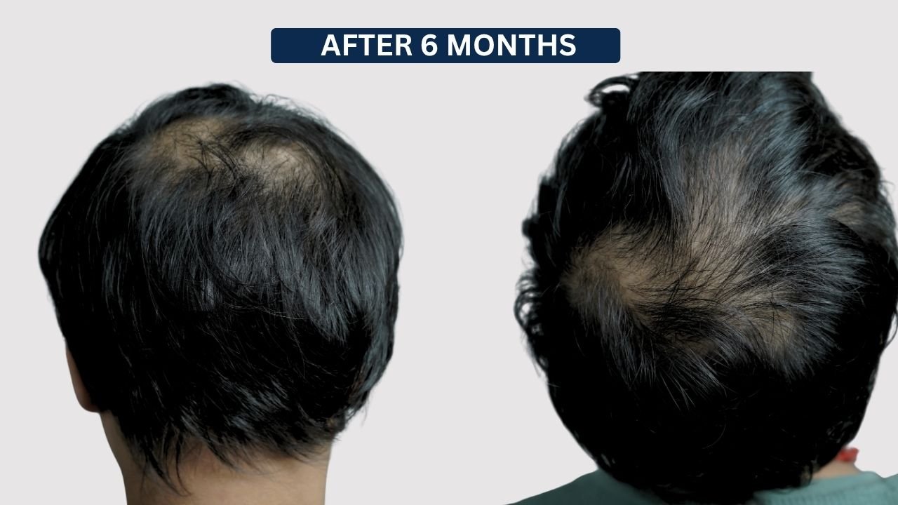
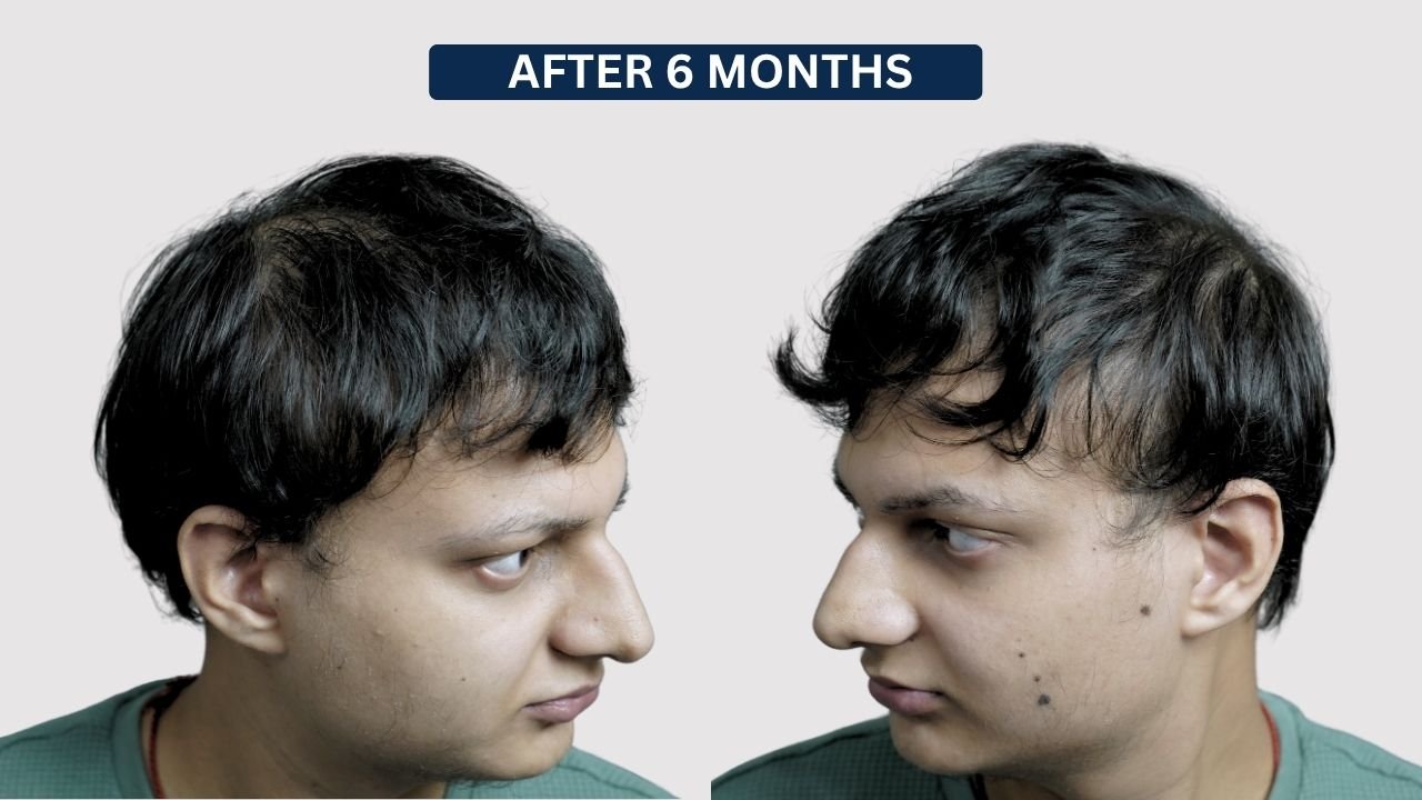
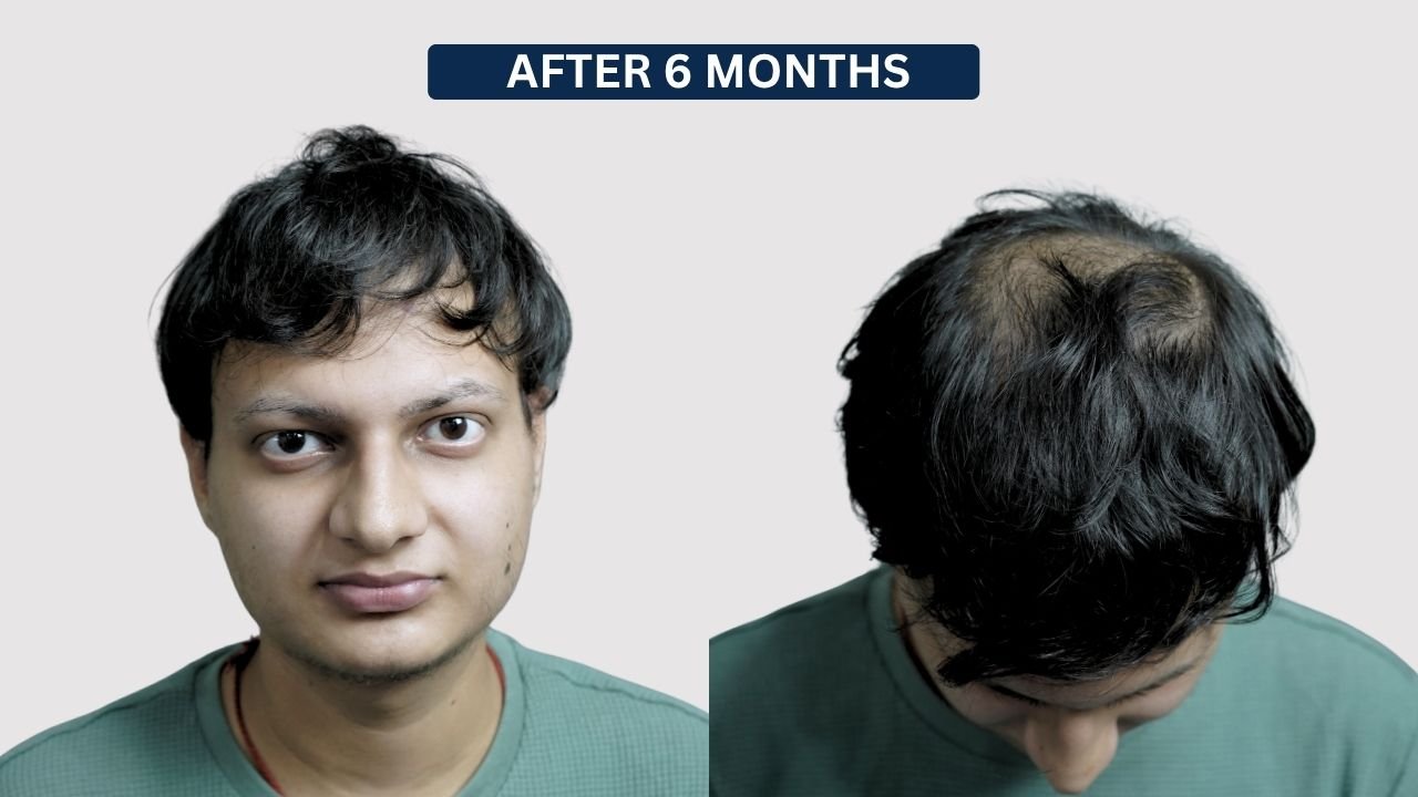
After 6 Months – Latest Documented Progress

At the 6-month follow-up, there was a clear visible improvement compared with the pretreatment condition.

The photographs demonstrate:

  • Considerably longer hair
  • Improved frontal scalp coverage
  • Better overall hair distribution
  • Increased visible hair density
  • Reduced scalp exposure
  • More mature hair growth compared with earlier stages
  • Improved cosmetic coverage of previously sparse areas

Some scalp visibility remained over the upper and crown regions when viewed from above, indicating that this was still an ongoing treatment stage rather than a guaranteed final result.

Treatment Progress Summary

Treatment Stage

Observed Progress

Before Treatment

Extensive scalp hair loss with marked scalp exposure and minimal visible hair

1 Month

Very early fine hair visible, but scalp remained largely exposed

3 Months

Developing regrowth with improved hair distribution

5 Months

Progressive increase in hair length and scalp coverage

6 Months

Latest documented stage with significantly better hair coverage and reduced scalp visibility

Patient Outcome & Observation

The patient’s serial photographs demonstrate progressive visible hair regrowth during the documented 6-month treatment journey.

At baseline, scalp exposure was extensive and only minimal visible hair remained.

During the first month, changes were limited, although fine hair could be seen in several areas. By three months, developing regrowth had become more noticeable.

Between the 3- and 5-month stages, the photographs show increasing hair length and broader scalp coverage.

By 6 months:

  • Overall hair coverage had improved significantly
  • Hair length had increased
  • Frontal coverage appeared more established
  • Scalp visibility was reduced
  • Regrowing hair appeared more mature
  • Residual thinning remained visible in some upper scalp areas

The 6-month photographs represent the latest documented progress, not necessarily the final outcome.

Aftercare & Long-Term Maintenance

Long-term dermatologist-directed follow-up remains important in alopecia areata because the condition can have an unpredictable course.

General care may include:

  • Follow the prescribed treatment schedule
  • Attend regular dermatologist follow-ups
  • Avoid unnecessary scalp irritation
  • Avoid aggressive chemical treatments when advised
  • Maintain gentle scalp and hair care
  • Monitor for sudden or localized new hair loss
  • Watch previously affected areas for renewed thinning
  • Report increased shedding to the treating dermatologist
  • Continue supportive treatment only if prescribed
  • Do not repeat, modify, or stop corticosteroid treatment without medical supervision

The supplied reference format likewise emphasizes continued monitoring and avoiding independent changes to prescribed corticosteroid treatment. Pasted markdown

The Change: A New Beginning

Long-term dermatologist-directed follow-up remains important in alopecia areata because the condition can have an unpredictable course.

General care may include:

  • Follow the prescribed treatment schedule
  • Attend regular dermatologist follow-ups
  • Avoid unnecessary scalp irritation
  • Avoid aggressive chemical treatments when advised
  • Maintain gentle scalp and hair care
  • Monitor for sudden or localized new hair loss
  • Watch previously affected areas for renewed thinning
  • Report increased shedding to the treating dermatologist
  • Continue supportive treatment only if prescribed
  • Do not repeat, modify, or stop corticosteroid treatment without medical supervision

The supplied reference format likewise emphasizes continued monitoring and avoiding independent changes to prescribed corticosteroid treatment. Pasted markdown

ATIYA SHAIKH Alopecia

What Our Patients Says

Why Choose Dr. Kiran Chotaliya for PRP Hair Treatment

  • Expert Hypertrophic Scar Care: Dr. Kiran Chotaliya provides personalized Kenacort Injection treatment plans based on the scar’s size, thickness, location, duration, symptoms, and overall skin condition.

  • Customized Scar Treatment Planning: The concentration, amount of medication, injection points, treatment interval, and number of sessions are decided according to the individual characteristics of the hypertrophic scar and the patient’s response.

  • Precise Intralesional Kenacort Injection: Kenacort is carefully injected directly into the raised scar tissue using a controlled intralesional technique to target the thickened area while minimizing unnecessary treatment of the surrounding skin.

  • Focus on Scar Flattening & Softening: Treatment is planned to help reduce the height, firmness, thickness, and prominence of the hypertrophic scar, while supporting a smoother and more even skin appearance.

  • Symptom-Oriented Scar Management: When hypertrophic scars are associated with discomfort, itching, tightness, or tenderness, the treatment plan may also focus on improving these symptoms based on the dermatologist’s clinical assessment.

  • Regular Follow-Up & Maintenance: Follow-up assessments help monitor changes in scar thickness, texture, softness, symptoms, and overall appearance. Further Kenacort sessions or additional scar-management options may be advised according to the patient’s progress and clinical response.

  •  
drkiranchotaliya

FAQ'S

1. What treatment was used in this alopecia areata case?

Kenacort contains triamcinolone, a corticosteroid. Intralesional corticosteroid injections may be used by dermatologists in selected alopecia areata cases, with dose, concentration, injection sites, and intervals individualized according to the patient.

Kenacort contains triamcinolone, a corticosteroid. Intralesional corticosteroid injections may be used by dermatologists for selected conditions, including localized alopecia areata and hypertrophic scars, although the technique, dose, and treatment plan differ according to the condition being treated.

Fine and short hair was visible during the 1-month follow-up, with more noticeable regrowth by approximately 3 months.

The photographs show a clearer improvement in overall coverage between approximately 3 and 5 months, with further visible improvement at 6 months.

Not necessarily. The 6-month stage is the latest documented follow-up for this case. Further changes may occur, and alopecia areata can recur.

Hair Transplant in Pune

Dr. Kiran Chotaliya is a leading hair transplant surgeon in Pune, specializing in advanced techniques like FUE and FUT for natural, lasting results. His expertise and patient-centered care ensure successful hair restoration with exceptional outcomes.

Follow Us

GET IN TOUCH

© 2026 Developed & Handle By Dr. Kiran Chotaliya Clinic Team

Location

© 2026 Developed & Handle By Dr. Kiran Chotaliya Clinic Team

Scroll to Top